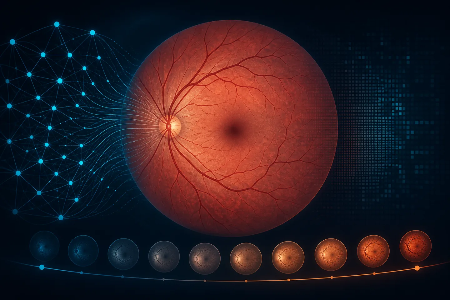
The retina exposes a network of blood vessels and neural tissue without an invasive procedure. Researchers fine-tuned the RETFound vision foundation model to estimate chronological age from colour fundus photographs. In 71,343 UK Biobank participants, mean absolute error reached 2.85 years.
The difference between predicted and actual age — the retinal age gap — was associated with cardiometabolic traits, inflammation, cognitive performance, all-cause mortality, dementia, cancer and incident cardiovascular disease. Genetic analyses pointed to pathways involving longevity, metabolism, neurodegeneration and age-related eye disease.
The team also found different biological patterns by sex: links to metabolic syndrome were stronger in males, while model attention and genetics pointed more strongly to retinal vasculature in females. Some female associations became more male-like after menopause. These are statistical relationships in observational data; the model does not establish causation and an individual score is not a life-expectancy forecast.
If the marker proves robust across populations and imaging systems, routine eye photography could eventually provide a low-cost signal for more focused prevention. Its realistic role would be triage — identifying people who may benefit from better cardiovascular, metabolic or cognitive assessment — rather than diagnosing disease from one photograph.
Translation requires prospective external validation, bias testing across ancestry, age and image quality, comparison with standard risk scores, and evidence that model-guided action improves care. Optimistically, validation pilots could appear in 2–4 years and limited clinical triage in 5–8 years if benefit is demonstrated. Broad screening cannot yet be dated.



Be the first to open the discussion.